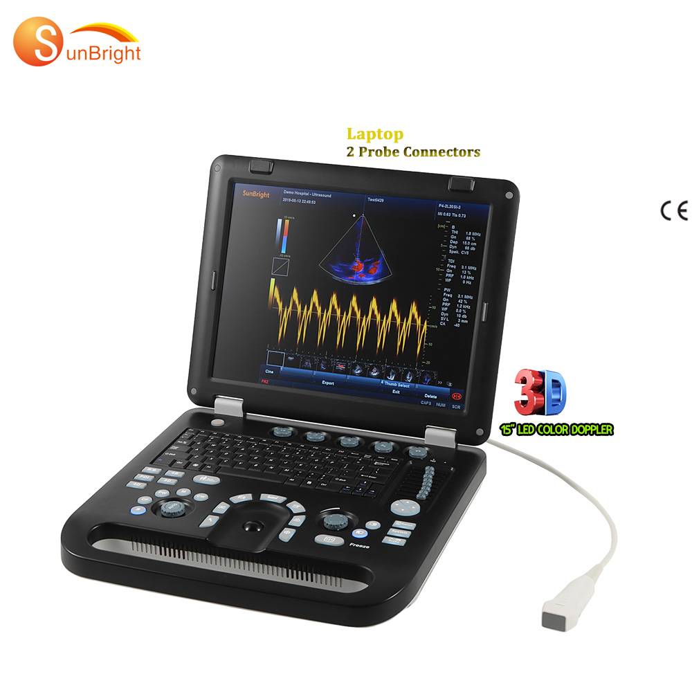
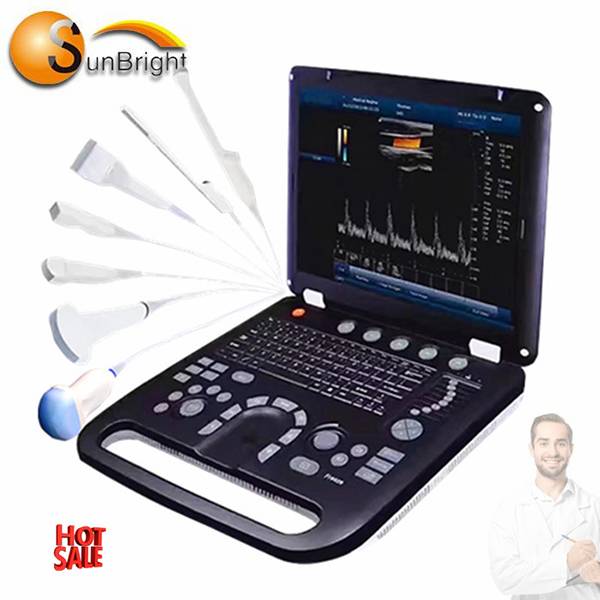
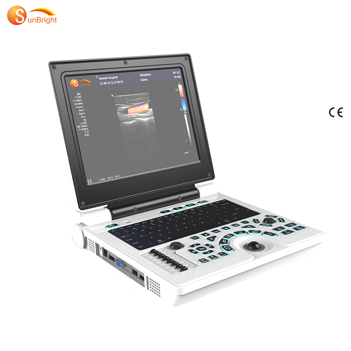
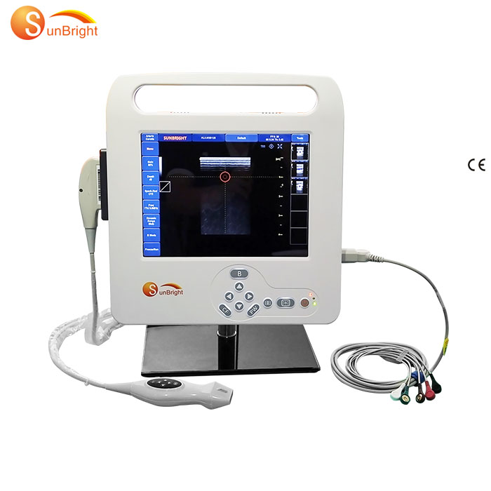
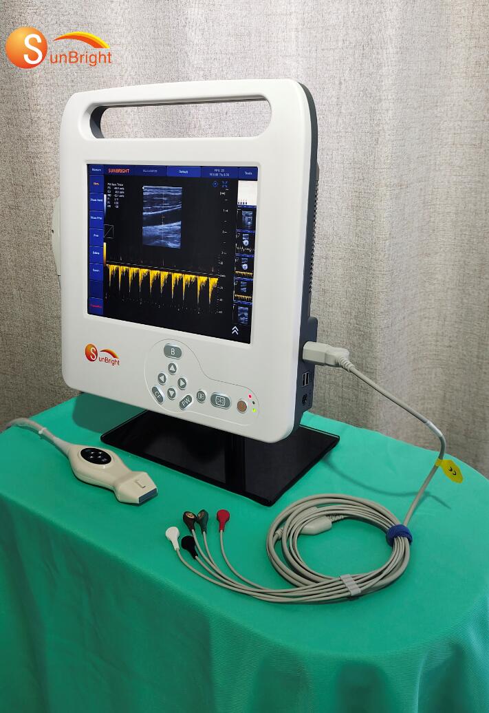
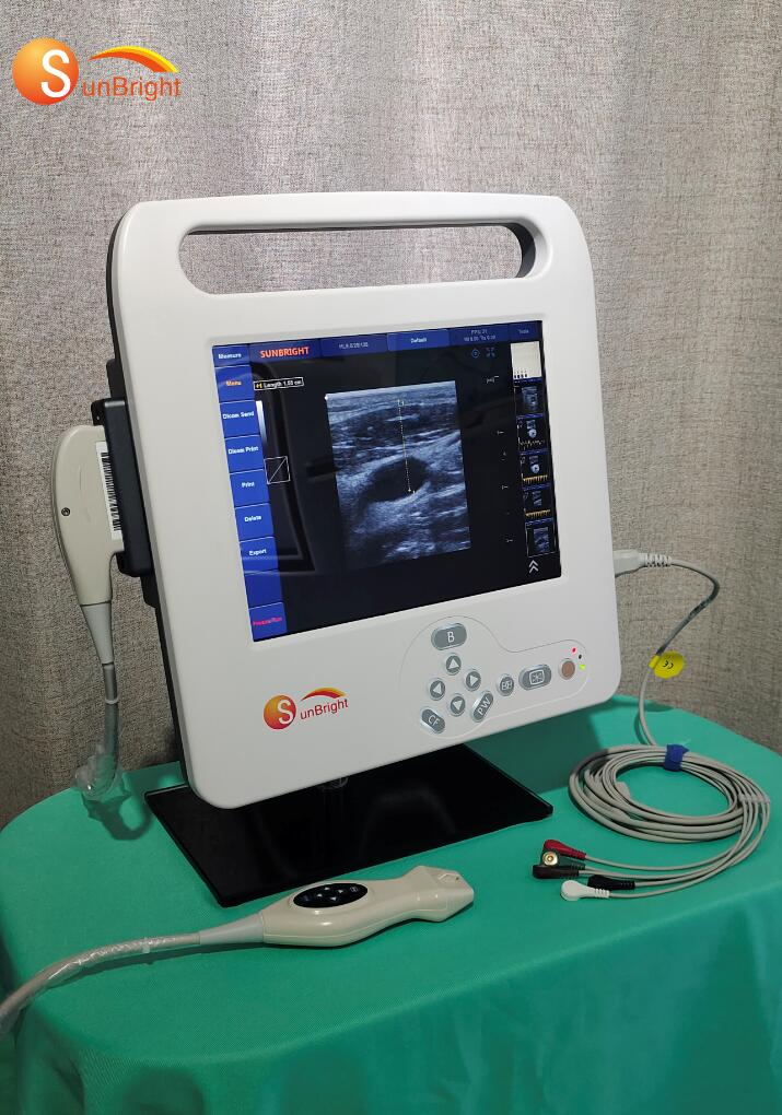
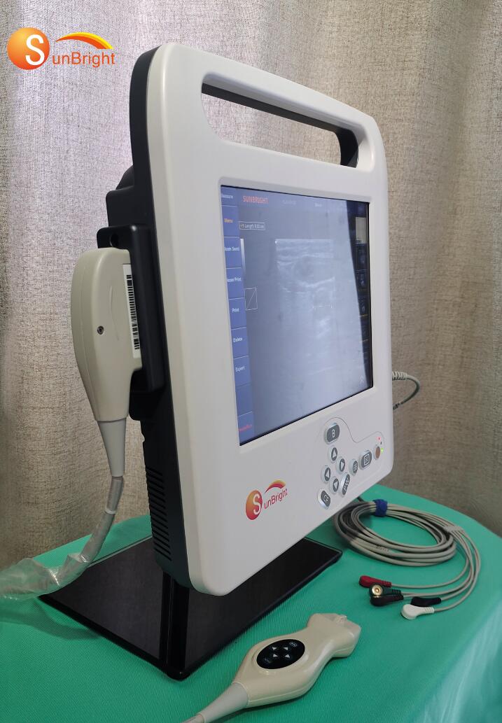
laptop high intensity medical CW diagnostic scanner color doppler SUN-906D


| Place of Origin | Shanghai, China |
| Brand Name | Sunbright |
| Model Number | SUN-906D |
| Instrument classification | Class II |
| Properties | Obstetric Appliances |
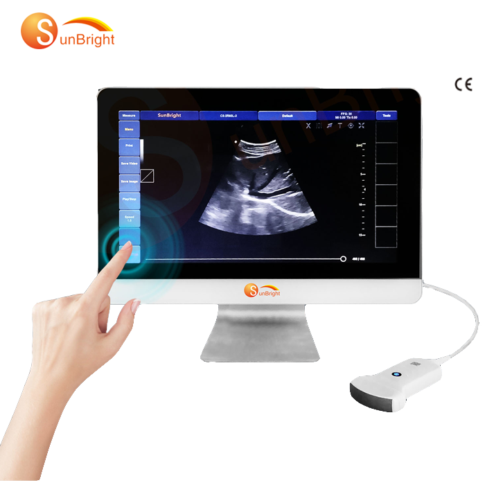
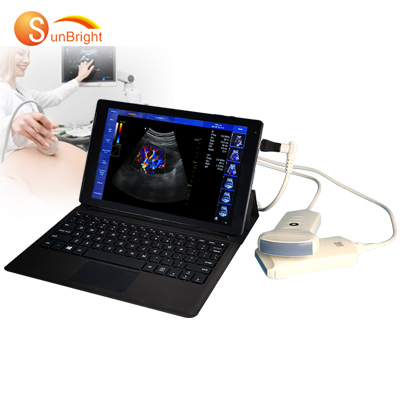
| Type | Clear image portable ultrasound |
| Scanning depth | 400mm max |
| Electrical Power supply | 100-240V~50/60Hz 100VA |
| Zoom | 10*magnify |
| size | 308mm*310mm*66mm |
| Net Weight | 4 Kg |
| Display | 12 inch LED |
| Resolution | 1024*768 |
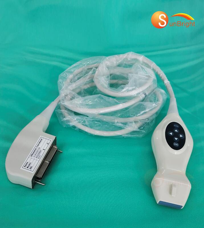
| Probe ports | 2-probe active ports |
| Hard disc | 120G SSD |
Supply Ability
Supply Ability:20000 Unit/Units per Year
Packaging & Delivery
Packaging Details:Sea-worthy packing/Air-worthy packing
Port:Shanghai
laptop high intensity medical color doppler CW diagnostic scanner
General Performance: | Digital Broadband | 12288 channels |
Beam-former | Re-programmable | |
Transmit Voltage | Adjustable (15 steps) | |
Beam-former Frequency Range | 1~40 MHz |
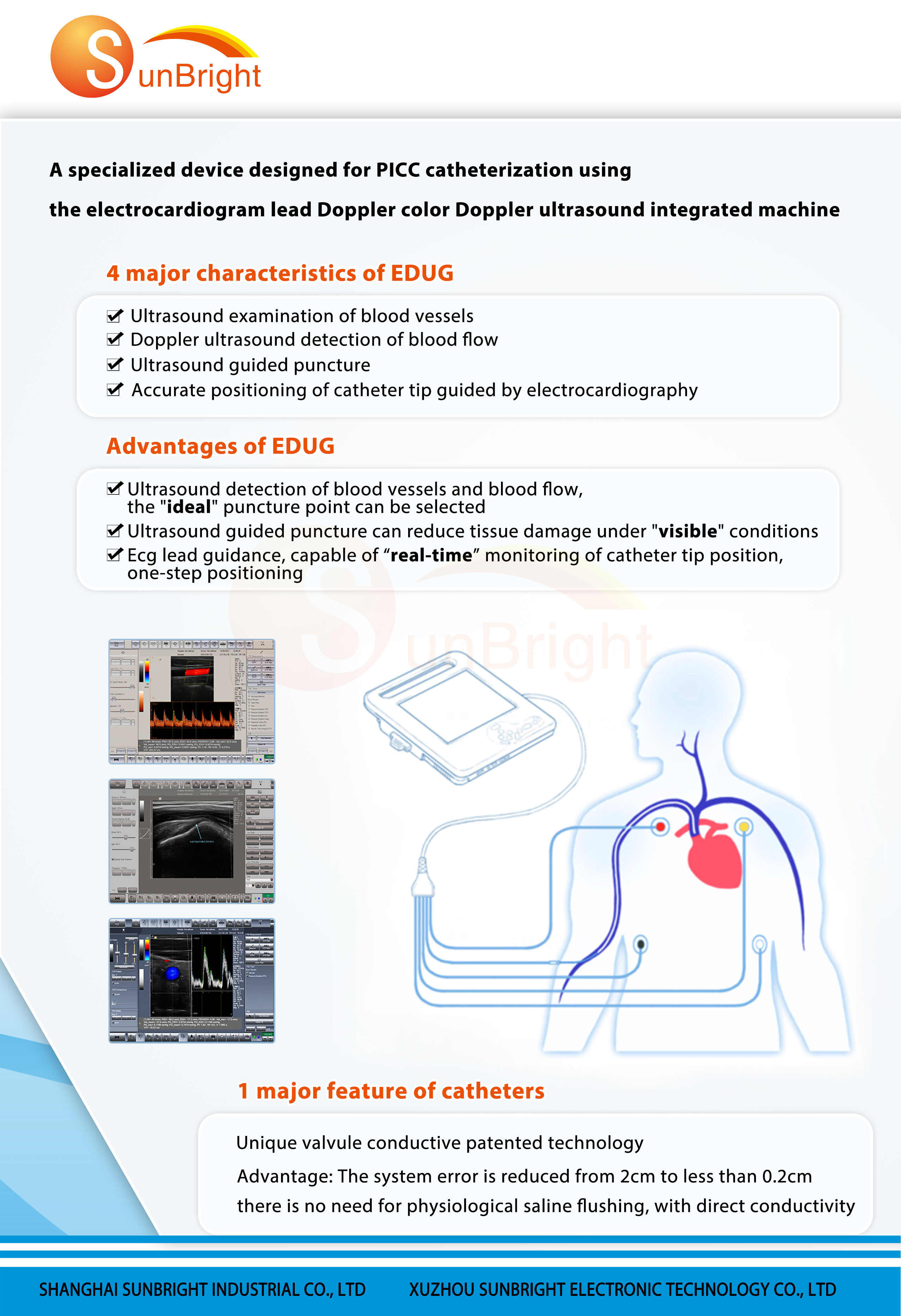
Doppler ultrasound detection of blood flow
Ultrasound guided puncture
Accurate positioning of catheter tip guided by electrocardiographyAdvantages of EDUG
Ultrasound detection of blood vessels and blood flow, the “ideal” puncture point can be selected
Ultrasound guided puncture can reduce tissue damage under “visible” conditions
Ecg lead guidance, capable of “real-time” monitoring of catheter tip position, one-step positioning
1 major feature of catheters
Unique valvule conductive patented technology
Advantage: The system error is reduced from 2cm to less than 0.2cm
there is no need for physiological saline flushing, with direct conductivity
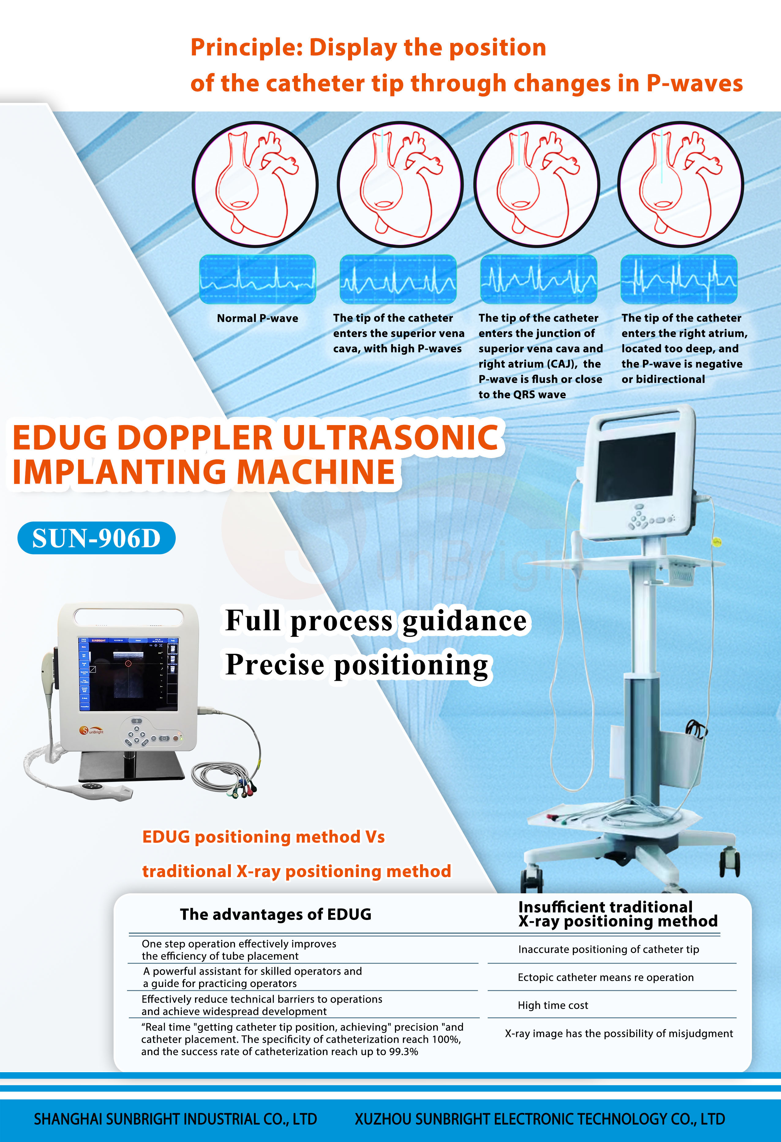
Principle; Display the position of the catheter tip through changes in P-waves
Normal P-wave
The tip of the catheter enters the superior vena cava, with high P-waves
The tip of the catheter enters the junction of superior vena cava and right atrium (CAJ), the
P-wave is flush or close to the QRS wave
The tip of the catheter enters the right atrium, located too deep, and the P-wave is negative or bidirectional

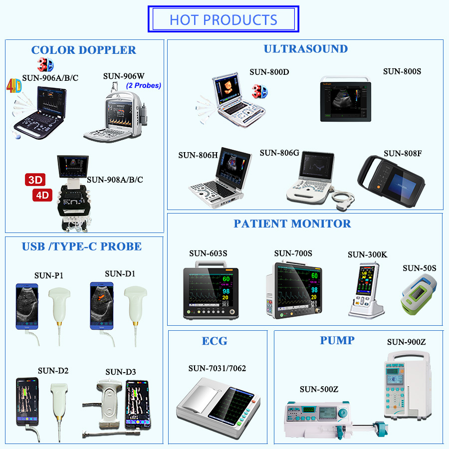
Recommend Products
Packing and delivery
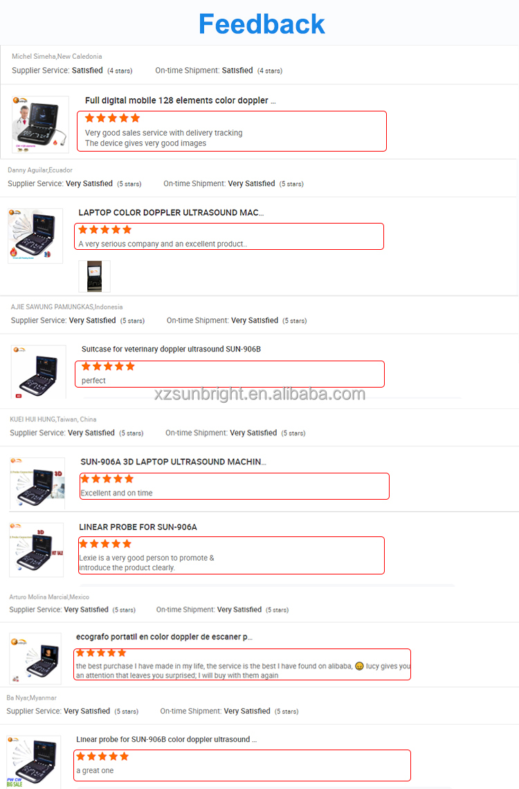
Why Choose Us
Certifications